The human eye is a complex organ that allows us to perceive the world around us. It relies on the process of refraction to focus light on the retina, which is located at the back of the eye. However, not all parts of the eye are involved in this refraction process. In this article, we will explore the anatomy of the eye, the process of light refraction, and the parts of the eye that do not play a role in focusing light on the retina.
Understanding the Anatomy of the Eye
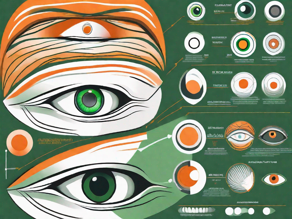
Before diving into the specifics of light refraction, let’s take a moment to understand the basic anatomy of the eye. The eye is a complex organ that allows us to perceive the world around us. It can be divided into several parts, each with its own unique function. These include the cornea, the lens, the iris, and the sclera.
The cornea, which is the transparent, dome-shaped front surface of the eye, acts as a protective barrier. It shields the delicate structures within the eye from external damage and helps to maintain the shape of the eye. In addition to its protective role, the cornea also plays a crucial role in the process of refraction.
The Role of the Cornea in Refraction
Refraction is the bending of light as it passes through different mediums. The cornea, being the first medium that light encounters when entering the eye, is responsible for the majority of the eye’s focusing power. It refracts, or bends, incoming light to help focus it on the retina, which is located at the back of the eye.
Imagine the cornea as a powerful lens that works in conjunction with the other parts of the eye to create a clear and focused image. Its curved shape and smooth surface allow it to bend the light in such a way that it converges on the retina, ensuring that the image we see is sharp and well-defined.
The Function of the Lens in Focusing Light
Located behind the cornea, the lens further refines the incoming light to ensure it is properly focused on the retina. The lens is a flexible and transparent structure that can change its shape to allow the eye to focus on objects at different distances.
When we look at objects that are far away, the lens becomes flatter, allowing light to be refracted less and enabling us to see distant objects clearly. Conversely, when we shift our focus to objects that are close to us, the lens becomes thicker and more curved, increasing its refractive power and allowing us to see nearby objects with clarity.
The ability of the lens to change its shape is known as accommodation. This process is controlled by the ciliary muscles, which surround the lens and contract or relax to alter its curvature. Accommodation is an automatic and continuous process that allows us to effortlessly shift our focus between objects at different distances.
By changing its curvature, the lens helps to fine-tune the refraction process and achieve clear vision. Together, the cornea and the lens work in harmony to ensure that light entering the eye is properly focused on the retina, where it is converted into electrical signals that are sent to the brain for interpretation.
The Process of Light Refraction in the Eye
Now that we understand the key components involved in the refraction process, let’s delve deeper into how light travels through the eye and the importance of focusing it on the retina.
How Light Travels Through the Eye
When light enters the eye, it first passes through the cornea, which bends and focuses the light. The cornea is a transparent, dome-shaped structure that covers the front of the eye. It plays a crucial role in the eye’s ability to refract light and helps protect the delicate structures within.
The light then continues its journey through the pupil, which is the small opening in the center of the iris. The iris, often referred to as the colored part of the eye, regulates the size of the pupil and controls the amount of light entering the eye. It acts like a camera aperture, adjusting its size based on the lighting conditions to optimize the amount of light reaching the retina.
After passing through the iris, the light encounters the lens, which further refracts the light and fine-tunes its focus. The lens dynamically adjusts its shape, allowing the eye to focus on objects at different distances, whether near or far. This process is known as accommodation and is controlled by the ciliary muscles surrounding the lens. When we focus on something close, the ciliary muscles contract, causing the lens to become thicker and more curved. Conversely, when we focus on something far, the ciliary muscles relax, making the lens thinner and less curved.
The Importance of Focusing Light on the Retina
At the back of the eye, we find the retina, which contains specialized cells called photoreceptors. The retina is like the film in a camera, capturing the focused light and converting it into electrical signals that can be interpreted by the brain. It is a complex network of cells, including rods and cones, that work together to process visual information.
For clear vision, it is essential that the light is accurately focused on the retina, as any inaccuracies can result in blurry or distorted vision. The cornea and lens work together to ensure that the light rays converge precisely on the retina, forming a sharp image. If the cornea or lens is misshapen or if the eye’s length is not ideal, it can lead to refractive errors such as myopia (nearsightedness), hyperopia (farsightedness), or astigmatism.
In conclusion, the process of light refraction in the eye is a remarkable feat of biological engineering. From the cornea to the lens to the retina, each component plays a crucial role in bending and focusing light to create clear and detailed vision. Understanding this process helps us appreciate the complexity and beauty of our visual system.
Parts of the Eye Not Involved in Light Refraction
While the cornea and lens play crucial roles in the process of light refraction, there are other parts of the eye that do not directly contribute to this process. Nonetheless, these parts still serve important functions in maintaining overall eye health and visual acuity.
The Iris and its Role in Eye Color
The iris, in addition to regulating the size of the pupil, determines eye color. The color of the iris is determined by the amount and distribution of pigmentation. While the iris does not directly participate in the refraction of light, it is an important part of the eye’s overall structure and function.
Interestingly, the iris is composed of two layers: the anterior and posterior layers. The anterior layer contains pigmented cells, while the posterior layer is made up of connective tissue. The combination of these layers gives the iris its unique color and pattern.
Eye color, which can range from shades of blue, green, brown, and even gray, is determined by the amount and type of pigmentation present in the iris. The more melanin pigment there is, the darker the eye color tends to be. However, it is important to note that eye color can also be influenced by other factors such as genetics and environmental factors.
In addition to its role in eye color, the iris also plays a crucial role in regulating the amount of light that enters the eye. It does this by adjusting the size of the pupil, which dilates or constricts in response to changes in lighting conditions. This mechanism helps to protect the delicate structures within the eye from excessive light exposure.
The Sclera and its Protective Function
Often referred to as the “white of the eye,” the sclera is the tough, fibrous outer layer that covers and protects the eyeball. It acts as a shield, providing structural support and preventing damage to the delicate internal parts of the eye. While the sclera does not contribute to light refraction, it plays a crucial role in maintaining the integrity of the eye.
The sclera is composed of dense collagen fibers that give it its strength and durability. It covers the entire surface of the eyeball, except for the cornea, and provides a protective barrier against external forces and potential injuries. Without the sclera, the eye would be vulnerable to damage from everyday activities such as rubbing or accidental impact.
In addition to its protective function, the sclera also serves as an attachment point for the extraocular muscles that control eye movements. These muscles, which are responsible for moving the eye in different directions, are anchored to the sclera. This allows for precise and coordinated movements of the eye, enabling us to focus on objects and navigate our surroundings.
Furthermore, the sclera also plays a role in maintaining the shape of the eye. Its rigid structure helps to keep the eyeball spherical, which is essential for proper focusing of light onto the retina. Any abnormalities or changes in the shape of the sclera can have a significant impact on visual acuity and overall eye health.
Overall, while the iris and sclera may not directly participate in the process of light refraction, they are integral parts of the eye’s anatomy and function. The iris determines eye color and regulates the amount of light entering the eye, while the sclera provides protection, attachment points for muscles, and helps maintain the shape of the eye. Understanding the roles of these structures enhances our appreciation for the complexity and intricacy of the human eye.
Common Eye Disorders Related to Light Refraction
Despite the intricate design of the eye, various factors can disrupt the process of light refraction, leading to vision problems. Some of the most common refractive errors include myopia, hypermetropia, and astigmatism.
Myopia and Hypermetropia: Issues with Light Focusing
Myopia, commonly known as nearsightedness, occurs when the eyeball is too long or the cornea is too curved. As a result, light focuses in front of the retina instead of directly on it, leading to blurry distance vision. This condition can make it difficult to see objects that are far away, such as road signs or classroom whiteboards.
Hypermetropia, on the other hand, occurs when the eyeball is too short or the cornea is too flat. In hypermetropia, light focuses behind the retina, causing difficulty in focusing on close objects. People with hypermetropia may experience eyestrain, headaches, and blurred vision when trying to read or perform tasks that require near vision.
Astigmatism: An Irregularity in Refraction
Astigmatism is a refractive error characterized by an irregularly shaped cornea or lens. Instead of being uniformly curved, the cornea or lens may have different curvatures in different meridians. This causes light to focus on multiple points on the retina, leading to blurry or distorted vision. People with astigmatism may have difficulty seeing both near and far objects clearly.
Astigmatism can occur in combination with myopia or hypermetropia, further complicating the visual impairment. It can cause visual discomfort, eye strain, and headaches. Individuals with astigmatism may also experience difficulties with night vision, as the irregularities in the cornea or lens can cause halos or glare around lights.
It is important to note that refractive errors can vary in severity. Some individuals may have mild myopia or hypermetropia that does not significantly impact their daily lives, while others may have more severe forms that require corrective measures such as glasses, contact lenses, or refractive surgery.
Correcting Refraction Errors
Refractive errors can be a hindrance to clear vision, but fortunately, there are various options available to correct them and improve visual acuity. The two most common methods used for this purpose are the use of glasses or contact lenses and refractive surgery.
The Role of Glasses and Contact Lenses
Glasses and contact lenses play a crucial role in correcting refractive errors. They work by altering the way light enters the eye, compensating for the specific refractive error present. By doing so, they provide a clear and focused image on the retina, allowing for improved vision.
When it comes to glasses, they are available in a wide range of styles and designs, catering to different preferences and needs. Whether it’s a trendy pair of frames or a more classic style, glasses offer not only vision correction but also an opportunity for self-expression and personal style.
Contact lenses, on the other hand, provide an alternative to glasses. They are small, thin lenses that are placed directly on the surface of the eye. Contact lenses offer the advantage of a wider field of vision and freedom from the frames of glasses. They are available in various types, including daily disposable, monthly disposable, and extended wear lenses, allowing individuals to choose the option that best suits their lifestyle.
Understanding Refractive Surgery Options
For those seeking a more permanent solution to refractive errors, refractive surgery is an option worth considering. Refractive surgery aims to correct refractive errors by reshaping the cornea, the transparent front part of the eye. By altering the cornea’s shape, the surgery improves its refractive properties, resulting in clearer vision.
One of the most well-known refractive surgery procedures is LASIK (Laser-Assisted In Situ Keratomileusis). LASIK involves creating a thin flap on the cornea using a microkeratome or femtosecond laser. The cornea is then reshaped using an excimer laser, which removes a precise amount of tissue. Finally, the corneal flap is repositioned, allowing for a quick recovery and minimal discomfort.
Another refractive surgery option is PRK (Photorefractive Keratectomy). PRK is similar to LASIK but does not involve creating a corneal flap. Instead, the outer layer of the cornea is gently removed, and the underlying tissue is reshaped using an excimer laser. Although the recovery time for PRK is longer compared to LASIK, it can be an excellent choice for individuals with thin corneas or other corneal irregularities.
SMILE (Small Incision Lenticule Extraction) is a newer refractive surgery technique that has gained popularity in recent years. It involves creating a small incision in the cornea and extracting a lenticule, a small piece of tissue, to reshape the cornea and correct the refractive error. SMILE offers the advantage of a minimally invasive procedure, potentially reducing the risk of certain complications associated with other refractive surgeries.
It’s important to note that not everyone is a suitable candidate for refractive surgery. Factors such as age, overall eye health, and the presence of certain eye conditions may affect eligibility. Therefore, a comprehensive eye examination and consultation with an experienced eye care professional are essential to determine the most appropriate treatment option.
Conclusion
In summary, the human eye is a remarkable organ that relies on the process of light refraction to focus incoming light on the retina. While the cornea and lens play a significant role in this process, other parts of the eye, such as the iris and sclera, serve important functions unrelated to refraction. Understanding the anatomy of the eye and the process of light refraction helps us appreciate the complexity of vision and the various methods available to correct refractive errors.
While taking care of your vision is essential, it’s just as important to nurture your overall well-being. The Vagus Nerve Stimulator by Vagus.net complements your health regimen by rewinding your autonomic clock. Experience the benefits of reduced stress, improved sleep, and a happier life with our advanced tVNS technology. Embrace a more balanced lifestyle today and Buy Now.